
MICROanatomy Kidney Model
Nursing Modals
₹82.00₹22.00
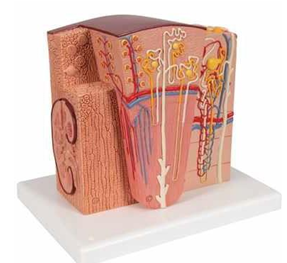
The microanatomy kidney model is a detailed representation of the intricate structures and functions of the kidney at the microscopic level. This model showcases various components such as nephrons, glomeruli, and renal tubules, allowing for an in-depth understanding of how these structures work together to filter blood and produce urine. It provides an excellent educational tool for students and professionals in the fields of medicine and biology, as it illuminates the complexities of renal physiology with clarity. By examining this model, one can appreciate the kidney's role in maintaining homeostasis, regulating electrolyte balance, and excreting waste products. Overall, the microanatomy kidney model serves as a valuable resource for enhancing knowledge about renal health and disease.
The Kidney is an extremely detailed model which shows the morphologic/functional units of the kidney. The kidney structures are greatly magnified. Six model zones illustrate the following fine-tissue structures of the human kidney that serve in the production of urine: Longitudinal section of a kidney Section of renal cortex and renal medulla of the kidney Wedge-shaped section of a kidney lobe with a diagrammatic depiction of three nephrons with Henle's loops of different lengths and diagrammatic depiction of the vascular supply Diagrammatic illustration of a kidney nephron with a short Henle's loop and didactic/diagrammatic illustration of the vascular supply Diagrammatic illustration of an opened kidney renal corpuscle with nephron and light-microscopic transverse sections of the proximal, attenuated and distal segments of a renal tubule Diagrammatic/didactic illustration of an opened kidney renal corpuscle MICRO Anatomy Kidney mounted on a base.